SINCHON·INT′l
SINCHON·INT′l
- MIRAE

show mobile menu
mobile menu
Array of hope: Up close and personal with mitochondria in neurons
Described as “the powerhouse of the cell,” mitochondria play a key role in cellular processes, and so, they are important to study when scientists investigate cell behaviour. Neurons or nerve cells depend on mitochondria to function properly, whether it be for managing membranes, or running processes for signal transmission and plasticity. Mitochondrial movement in neurons defines the health of the neurons, as these “powerhouses” provide vital energy to neurons. Due to their cellular responsibilities, mitochondria must be distributed appropriately in a neuron network. Over 30% of mitochondria in a healthy neuron are moving. Understanding their distribution and movement is therefore important.
Traditionally, methods like surface plasmon microscopy have been used to analyze mitochondrial movement in cells. Now, researchers from Yonsei University, in Seoul, Korea, have investigated movements in neuronal mitochondria via imaging techniques that are said to be more accurate than traditional methods, in their study recently published in ACS Nano. Prof Donghyun Kim and the rest of the team tracked mitochondrial movement across 3D space in real time by utilizing localized light beams created by plasmonic nanohole arrays (PNAs).
 Neurons transmit electrical signals across intricate networks, while mitochondria help to manage processes on the cellular level.
(Photo courtesy: Freepik)
Previously, traditional imaging methods showed only a partial picture of mitochondrial movement in neurons. Gaps remained in the puzzle of mitochondrial distribution and its implications for disease development. A full picture of mitochondrial dynamics has been sought after, as Prof Kim says, “an analytical method capable of three-dimensional tracking and imaging, which allows more precise and quantitative measurement of mitochondrial movement in live cells, is highly desired.”
To achieve this desired method, the research team used Plasmonic Nanohole Arrays. Fluorescence excited by the PNAs was captured with an optical microscope, where superlocalized light beams provided the required precision. This technique offered exceptional optical transmission via the PNAs, to follow mitochondrial movement at a 3D level. Higher 2D resolution wasn’t necessarily achieved, but the method offers much higher precision in capturing the movements of mitochondria, especially with light fields being highly localized. This high precision 3D tracking is a vast improvement over previously used microscopy techniques. Real-time analysis of neurons in 3D provided a resolution enhanced by 12.7 fold compared to the traditional approach of confocal fluorescence microscopy. This method is therefore superior to existing imaging methods for nanostructures, and can thus be used for highly reliable imaging of several cellular nanostructures.
This study successfully revealed mitochondrial movements at 50% velocity in the “depth” axis. PNAs provided depth information, allowing for a 3D interpretation of mitochondrial distribution. These details can help trace neuron structures, even down to the nanoscale, for objects like receptors. The team suggested significant movement on the depth axis could reflect a network of microtubules within a neuronal “axon”—an extension that carries electrical signals away from a neuron’s cell body.
As Prof Kim concludes, “mitochondrial positions were acquired with superlocalized precision.” Arrays of light beams superlocalized by PNAs helped track mitochondrial movements in neuron cells with high precision. Deeper insights into mitochondrial movement, like those found in this study, are valuable for understanding cellular functions and even disease development.
Updated in July 2019
Recommended Articles
Professor Jong-Hyun Ahn
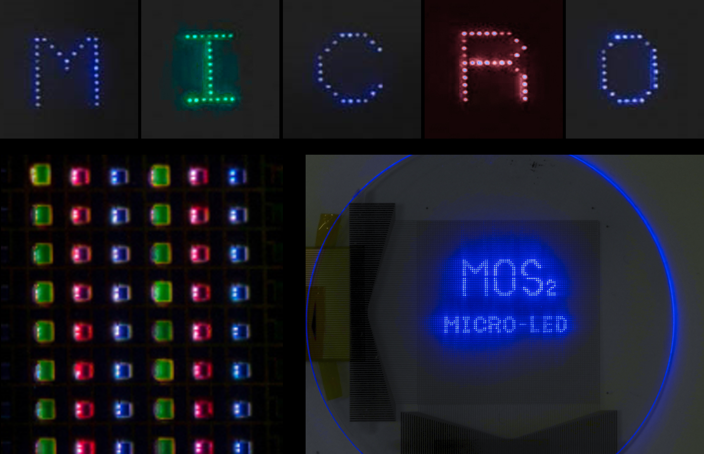
Novel technique for producing high-resolution micro-LED displays
Professor Ki Jun Yu
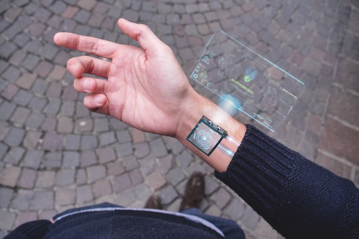
‘Skintilla’ of Warmth: A Breakthrough in Wearable Body Temperature Monitoring

Professor Seong Chan Jun


