SINCHON·INT′l
SINCHON·INT′l
- MIRAE

show mobile menu
mobile menu
Smarter MRI Diagnosis with Nano MRI Lamp
IBS scientists devise a new platform to overcome the limits of MRI contrast agents.
A research team led by Jinwoo Cheon (Department of Chemistry, Yonsei University) at the Center for Nanomedicine, within the Institute for Basic Science (IBS), developed the Nano MRI Lamp: a new technology platform that tunes the magnetic resonance imaging (MRI) signals “ON” only in the presence of the disease target. Published in Nature Materials, this study can overcome the limitations of existing magnetic resonance imaging (MRI) contrast agents.
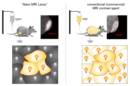
MRI is an increasingly popular non-invasive technique for diagnosis, which does not use harmful radiation. Some tissues show a natural contrast on MRI, but for some specific types of imaging, patients are administered an MRI contrast agent, to enhance the difference between the target area and the rest of the body. “Typical MRI contrast agents, like the most commonly used one, gadolinium, are injected in “ON” state and distributed across the whole biological system with relatively large background signal. Cheon explains, “We found a new principle to switch the MRI contrast agent “ON” only in the location of the target”. IBS scientists discovered how to switch the signal ON/OFF by using the Nano MRI Lamp.
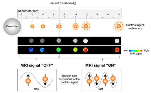
The Nano MRI Lamp consists of two magnetic materials: a quencher (magnetic nanoparticle) and an enhancer (MRI contrast agent). The switch is due to the distance between the two. When the two materials are at farther than a critical distance, 7 nanometers, the MRI signal is “ON”, whereas when they are placed closer than 7 nanometers, the MRI signal is “OFF”. The researchers named this phenomenon Magnetic REsonance Tuning (MRET), which is analogous to the powerful optical sensing technique called Fluorescence Resonance Energy Transfer (FRET).
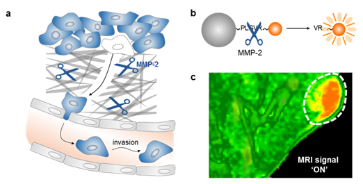
The researchers tested the Nano MRI Lamp for cancer diagnosis. They could detect the presence of an enzyme that can induce tumor metastasis, MMP-2 (matrix metalloproteinase-2) in mice with cancer. They connected the two magnetic materials with a linker that is naturally cleaved by MMP-2. Since the linker keeps the two materials close to each other, the MRI signal is “OFF”. However, in the presence of the cancer, the linker is cleaved by MMP-2, which cause the two materials to be separated and the MRI signal turns “ON”. Therefore, the MRI signal indicates the location of MMP-2, and so of the tumor. The scientists also found that the brightness of the MRI signal correlates with the concentration of MMP-2 in the cancerous tissue.
Most importantly, the Nano MRI Lamp is off until it meets a biomarker associated with a specific disease, allowing higher sensitivity. “The current contrast agent is like using a flashlight during a sunny day: its effect is limited. This new technology, instead, is like using a flash light in the night: it’s more useful,” exemplifies Cheon.
Beyond cancer diagnosis, in principle, the Nano MRI Lamp can be applied to investigate a variety of biological events, such as enzymolysis, pH variation, and protein-protein interactions, and so on. IBS scientists expect that it would be useful for both in vitro and in vivo diagnostics.
“Although we still have a long way to go, we established the principle and believe that the MRET and Nano MRI Lamp can serve as a novel sensing principle to augment the exploration of a wide range of biological systems,” concludes Cheon. The research group is now working on developing safer and smarter multitasking contrast agents, which can simultaneously record and interpret multiple biological targets, and eventually allow a better understanding of biological processes and accurate diagnosis of diseases.
Recommended Articles

Professor Byeong-Su Kim
New study demonstrates that “deformable” electronics are not a stretch

Professor Yeonjin Yi


